
Central Pressure Waveform Animations
The arterial system is directly responsible for moderating forward and reflected wave interactions and therefore the shape of the central pressure waveform. The following animations show how changes to the state of the arteries, sites of wave reflections, and age all contribute to changes in the contour of the central pressure wave.
Incident and reflected waves
The beginning of the incident pressure wave generated by the heart (blue) is shown traveling down the artery. When the wavefront encounters a change in vessel impedance (e.g., a change in resistance), a reflected wave (red) is generated and travels back toward the heart.
In the arterial system, resistance remains relatively constant through the large arteries, but increases significantly at the level of the arterioles. Thus, the arterioles represent the major site of origin of the reflected wave.
These incident and reflected waves combine to produce the pressure at any point in the arterial system.The pressure waveform sensed by a transducer (and the arterial wall) at point A is shown. The magnitude and shape of the combined pressure wave depend on both the speed at which the waves travel up and down the arteries (the pulse wave velocity) and the strength of the reflected wave.
Peripheral vasoconstriction produces only a slight increase in the amplitude of the reflected wave (since peripheral resistance is already quite high). On the other hand, peripheral vasodilation reduces the amplitude of the reflected wave and delays its return. A stiffer aorta produces an increase in amplitude of the incident wave along with a stronger reflected wave that returns earlier.
Arterial reflection sites
Every impedance mismatch along the arterial tree contributes to the reflected wave. However, the primary source for reflection is in the arterioles. The reflected waves from the many arterioles in the trunk and lower limbs coalesce to provide what appears to be a single reflected wave from an average or functionally discrete reflecting site.
Note that the reflected wave from the abdomen and lower limbs travels back to the the upper limbs.
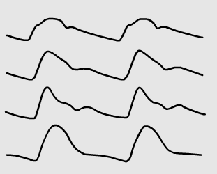
Aortic pressure changes with age
The central aortic pressure waveform is shown with its component waves from the second to the eighth decades.
The incident wave becomes progressively greater with age. The reflected wave increases to a similar degree, and returns progressively earlier.